Neuropathic pain is a difficult task for attending physicians. Problems with its treatment can be associated with various factors, including a complex clinical picture, the wrong choice of a drug and its dose. Not all patients can name their sensations characteristic of this pathological condition, which can lead to an incorrect diagnosis, especially by general practitioners, who are referred by most patients with neuropathic pain. This type of pain is difficult to treat, and few patients manage to completely stop it. As a rule, patients have sleep disturbance, depression and anxiety develop, and the quality of life decreases. Many of them suffer for a long time before receiving adequate assistance. Most patients (about 80%) experience pain for more than a year before their first visit to a specialist.
Another factor is the wrong drug choice. A key characteristic of neuropathic pain is that it does not respond well to traditional pain medications, such as non-steroidal anti-inflammatory drugs. Anticonvulsants and antidepressants are considered the most effective drugs for its treatment. However, as practice shows, these drugs make up only a small part of all medical prescriptions (about 20%) for neuropathic pain. While non-steroidal anti-inflammatory drugs for neuropathic pain are prescribed in 41% of cases, simple analgesics in 21%. Thus, more than 60% of patients with neuropathic pain receive inadequate pharmacotherapy!
Nevertheless, in recent years significant progress has been made in understanding the mechanisms of neuropathic pain and new possibilities for its effective therapy have appeared.
Terminology. From the standpoint of pathophysiology, it is now customary to distinguish between nociceptive and neuropathic pain. The term "nociceptive" comes from lat. here - to damage. Nociceptive pain occurs when a tissue damaging stimulus acts on peripheral pain receptors (nociceptors) located in various tissues of the body (skin, smooth and striated muscles, ligaments, joints, capsules of internal organs, etc.). An exogenous nociceptor can be both exogenous mechanical, thermal factors, and endogenous processes (inflammation, muscle spasm). Nociceptive pain is most often acute and has a protective function (“pain is the watchdog of health” by C. Sherington). This is a warning signal of danger, which forces a person to take appropriate measures. An example of nociceptive pain can be pain from a burn, bruise, trauma, acute or chronic inflammatory process, muscle spasm, myocardial infarction, etc. With this pain, the pain causing factor is usually obvious, the pain is usually accurately localized and easily described by patients. It should be emphasized that the conduct of the pain signal along the nervous system is not disturbed. An important aspect of nociceptive pain is that it induces the so-called antinociceptive endogenous systems (analgesic), as a result of which, in most cases, acute nociceptive pain quickly subsides or completely disappears. Antinociceptive systems have a modulating effect on all stages of the pain signal and its perception, determining individual pain response and behavior. Characteristic of nociceptive pain is its rapid regression after the termination of the damaging factor and after the use of painkillers.
Neuropathic refers to pain arising from an organic lesion or impaired function of various parts of the nervous system involved in pain control. It can be caused by damage to the nervous system at any level, from peripheral sensory nerves to the cerebral cortex. In neurology, the term "neuropathic" or "neuropathic" is usually understood as damage to the peripheral nerve. In this regard, it is possible that neuropathic pain is pain exclusively with peripheral neuropathy or polyneuropathy, so it should be emphasized once again that the term "neuropathic pain" characterizes pain in case of damage or impaired function of both the peripheral and central nervous system on any level.
Nociceptive pain is more often acute, less often chronic (osteoarthritis, rheumatoid arthritis). Neuropathic pain is predominantly chronic.
The choice of pharmacotherapy depends on the correct interpretation of the pain syndrome (nociceptive, neuropathic or mixed). With nociceptive pain, non-steroidal anti-inflammatory drugs are recommended, which have proven effective in this type of pain syndrome. In case of neuropathic pain, drugs of a different mechanism of action (anticonvulsants, antidepressants, opioids, etc.) are indicated, which will be discussed in the "treatment" section. Thus, for adequate therapy, analysis of the pain syndrome from the point of view of pathophysiological mechanisms is very important.
Epidemiology. According to American studies, neuropathic pain occurs in 1.5% of cases. In Europe, its prevalence is 6.5-7.5% in the population. Today, the concept of neuropathic pain unites a large group of chronic pain syndromes that were previously considered independently.
Peripheral neuropathic pain, Diabetic polyneuropathy, Alcoholic polyneuropathy, Chemotherapy-induced polyneuropathy, Acute and chronic inflammatory demyelinating polyneuropathy, Alimentary-mediated polyneuropathy, Idiopathic sensory neuropathy, Compression or infiltration, neurologic neuropathy, neuralgia pain neuropathy, Tunnel neuropathy, Radiculopathy (cervical, lumbosacral), Pain after mastectomy, Post-radiation plexopathy, Complex regional pain syndrome, Central neuropathic pain, Compression myelopathy with spinal canal stenosis, Post-radiation myelopathy, Vascular myelopathy, HIV-related myelopathy, Spinal cord injury, Post-stroke pain, Pain with multiple sclerosis, Sclerosis sclerosis, Sclerosis.
Lateral brain stem stroke
This group includes pain with various mono- and polyneuropathies, most often pain occurs with diabetic and alcoholic polyneuropathy (25–45%). Postherpetic neuralgia (in old age this complication occurs in 70% of cases of herpes zoster) is also a variant of neuropathic pain. Neuropathic pain syndrome includes a complex regional pain syndrome (local pain with edema, trophic disorders and osteoporosis), which was previously designated as reflex sympathetic dystrophy. Trigeminal neuralgia, phantom pain, post-stroke central pain, pain with multiple sclerosis, syringomyelia, spinal cord injury are typical examples of neuropathic pain. According to various authors, it occurs: in diabetic polyneuropathy - up to 45%, multiple sclerosis - 28%, syringomyelia - 75%, cerebral stroke - 8%, nerve injury - 5%. Most patients with neuropathic pain (about 50%) are elderly people (radiculopathy, diabetic polyneuropathy and postherpetic neuralgia). Despite the fact that in recent years they began to consider radiculopathy as a variant of neuropathic pain, it should be noted that it is not exclusively neuropathic in its mechanism. It is more correct to talk about the participation of both neuropathic and nociceptive components in the formation of this pain, since in addition to the direct damage to the nerve root, radiculopathy activates the nociceptors of the intervertebral disc, ligamentous apparatus, and a nonspecific inflammatory process develops.
The clinical characteristic. Neuropathic pain has its own characteristic differences. First of all, it is a complex of various sensory disorders. Specific to neuropathic pain is the phenomenon of allodynia. Allodynia is the appearance of pain in response to a stimulus that, under normal conditions, does not cause it. In such cases, patients experience severe pain at the slightest touch, sometimes even when the wind blows. Distinguish between temperature (the effect of a temperature stimulus) and mechanical (the effect of a mechanical stimulus) allodynia. Mechanical allodynia is divided into static, which appears when pressure is applied to a fixed point on the skin, and dynamic, which occurs when moving stimuli, such as mild skin irritation with a brush or finger.
With neuropathic pain, hyperesthesia, hyperalgesia, hyperpathy, neuralgia are often observed. The term “hyperalgesia” is used if the sensitivity, which under normal conditions was supposed to cause pain, was much higher than expected. According to localization, primary and secondary hyperalgesia is distinguished. Primary hyperalgesia is localized in the innervation zone of the damaged nerve or in the zone of tissue damage. Secondary hyperalgesia is more widespread, going far beyond the boundaries of tissue damage or the innervation zone of the damaged nerve. With hyperpathy, the subjective response to both pain and non-pain stimulus is excessive and often persists for a long time after the cessation of irritation. The phenomenon of neuralgia (trigeminal, postherpetic) is a typical example of neuropathic pain.
The patient may also be disturbed by spontaneous pain arising from the apparent absence of any external effects. She, as a rule, burning, stitching. Feeling of tickling, painless tingling or other similar sensations belong to paresthesia; if these sensations cause pain, they are called dysesthesia.
General characteristic of neuropathic pain:
- persistent (permanent) character;
- long duration;
- ineffectiveness of analgesics for its relief;
- diverse sensory involvement, i.e., positive sensitive phenomena: paresthesia, dysesthesia, neuralgia, hyperesthesia, hyperalgesia, allodynia, or negative symptoms (loss of different sensitivity modalities - hypalgesia, thermohypesthesia);
- a combination with vegetative symptoms;
- combination with motor disorders.
Diabetic polyneuropathy. Pain syndrome in diabetic polyneuropathy is a typical example of neuropathic pain with a combination of positive and negative sensory phenomena, most often in patients with distal symmetric sensory and sensorimotor polyneuropathy. Typical complaints are tingling and numbness in the feet and legs, worse at night. At the same time, patients may experience a sharp, shooting (lancing), throbbing and burning pain. Some patients have allodynia and hyperesthesia. All of the above disorders are classified as positive sensory symptoms of neuropathic pain. Negative symptoms include pain and temperature hypesthesia, which in the initial stage of the disease is moderately presented in the distal section, but as it progresses, it spreads proximally, involving the hands. Tendon reflexes are usually reduced, and muscle weakness is limited to the muscles of the foot.
Less commonly, pain can occur in diabetic asymmetric neuropathy due to vasculitis in epineuria in elderly people with mild or undetermined diabetes mellitus. Pain often begins in the lumbar region or in the hip joint and extends distally along the leg on one side. In this case, weakness and weight loss of the muscles of the thigh and pelvis on the same side are noted. Recovery is generally good, but not always complete.
Diabetic thoracolumbar radiculopathy due to damage to the nerve roots is characterized by pain in the shoulder girdle on one or both sides, in combination with skin hyperesthesia and hypesthesia in the area of the affected roots. This form is more common in elderly patients with long-term diabetes mellitus and, as a rule, tends to slow recovery of functions.
In severe violation of carbohydrate metabolism (ketoacidosis), acute neuropathy with severe burning pain and weight loss is possible. Allodynia and hyperalgesia are very pronounced with minimal sensory and motor deficits. Recovery correlates with weight gain and glycemic correction.
Postherpetic neuralgia is the most common complication of herpes virus infection, especially in the elderly and in patients with impaired immunity. Herpes zoster, or herpes zoster, is an acute painful condition. After varicella chickenpox, transmitted in childhood, the Varicella zoster virus remains latent in the body, localizing mainly in the sensory ganglia located in the posterior roots of the spinal nerves and in the sensitive roots of the trigeminal nerve. During reactivation (including sometimes after vaccination), the virus causes the formation of a characteristic vesicular rash located in the dermatome type, i.e. in the innervation zone of the corresponding sensory nerve: in 50% of patients on the trunk, in 20% on the head, in 15% - on the hands and 15% - on the feet. After a few days, the rash transforms into a pustular, then forms crusts and disappears by the end of the 3rd week of the disease.
Although in some patients the acute phase of herpes zoster is asymptomatic, in most cases postherpetic neuralgia develops. This complication is observed in 50% of patients older than 60 years. The pain associated with such neuralgia is based on inflammatory changes or damage to the ganglia of the posterior roots of the spinal cord and peripheral nerves in the affected areas of the body. If the posterior root or its ganglion is affected, pathological changes also occur in the corresponding innervation zone. Postherpetic neuralgia occurs with damage to the roots of the posterior horns of the spinal nerves, most often thoracic, and trigeminal nerves.
Most often postherpetic neuralgia persists:
- within 1 month after the rash appears,
- within 3 months after the rash appears,
- after the disappearance of the rash.
Postherpetic neuralgia may be:
- constant, deep, dull, oppressive or burning,
- spontaneous, periodic, stitching, shooting, similar to "electric shock",
- allodynic, acute, superficial, burning, radiating, itchy when dressing or lightly touching (in 90% of patients).
In most patients, postherpetic neuralgia decreases during the first year. However, in some patients, it can persist for years and even throughout the rest of their lives, causing considerable suffering. Neuralgia has a significant negative impact on the quality of life and the functional status of patients who may develop affective disorders in the form of anxiety and depression.
Central post-stroke pain
The term "central post-stroke pain" refers to pain and some other sensory disturbances resulting from a stroke, unless other causes of pain are identified. This pain is also called thalamic, although it has now been established that cerebrovascular accidents in other parts of the central nervous system, in addition to the optic tubercle, can cause central neuropathic pain. In general, central neuropathic pain is observed in approximately 8% of stroke patients. Nevertheless, the visual tubercle and brain stem are parts of the brain, the defeat of which during a stroke is most often accompanied by central neuropathic pain. Her pathophysiology remains largely unclear.
Central post-stroke pain develops in the first year after a stroke in 8% of patients. Since the prevalence of stroke is high - about 500 cases per 100,000 population - the absolute number of people with post-stroke pain is very significant. The onset of pain can be soon after a stroke or after a certain time. In a special study of this pain, the following data were obtained: in 50% of patients, pain occurred during the first month after a stroke, in 37% it began in the period from a month to 2 years after a stroke, in 11% - after 2 years from the moment of a stroke.
Central post-stroke pain is felt in a large part of the body, for example, in the right or left half; however, in some patients, pain may be localized in one arm or in the face. Most often they characterize it as burning, aching, plucking, tearing. Various factors can exacerbate post-stroke pain: movement, cold, warmth, emotions. In other patients, by contrast, these same factors can alleviate pain, especially heat. This pain is often accompanied by neurological symptoms such as hyperesthesia, dysesthesia, numbness, changes in sensitivity to heat, cold, touch and / or vibration. Pathological sensitivity to heat and cold is most common and serves as a reliable diagnostic sign of central neuropathic pain. According to studies, 70% of patients with this pain are not able to feel the difference in temperature in the range from 0 to 50 °. It should be noted that pain is very difficult to treat and often refractory to opiate therapy, tricyclic antidepressants and carbamazepine.
Diagnostics. The key to the diagnosis of neuropathic pain syndrome is often in neglected or completely forgotten events that occurred several weeks or months before the onset of symptoms of the disease. Therefore, the patient should be carefully questioned about recent infections, injuries, to find out if he has any other symptoms of a systemic disease, endocrine pathology, and should be asked about the patient's attitude to alcoholic beverages. It is important to find out how the disease started, as the first symptoms, such as paresthesia or numbness, may appear several days or even weeks before the onset of pain. Much attention should be paid to the characteristic given by the patient to the most pain syndrome: intensity, duration, variability during the day; factors that can provoke it, as well as the effectiveness of analgesics.

During clinical and neurological examination, it is important to focus on the condition of not only the sensitive sphere, identifying positive (allodynia, hyperalgesia, hyperpathy) and negative (hypalgesia) symptoms, but also motor (peripheral paresis, atrophy, central paresis, spasticity, hyperkinesis), as well as autonomic nervous system (violation of sweating, swelling, discoloration of the skin).
Of the scales designed to assess neuropathic pain, the most popular are:
DN4 questionnaire for diagnosing the origin of neuropathic pain.
Please complete this questionnaire by ticking one answer for each item in the 4 questions below.
Patient Interview
Question 1: Does the pain experienced by the patient correspond to one or more of the following definitions? (Well no)
- Burning sensation
- Painful sensation of cold
- Sensation of electric shock
Question 2: Is pain accompanied by one or more of the following symptoms in the area of its localization? (Well no)
- Pinching, crawling sensation
- Tingling
- Numbness
- Itching
Patient examination
Question 3: Is the pain localized in the same area where the examination reveals one or both of the following symptoms: (Yes / No)
- Reduced touch sensitivity
- Reduced tingling sensitivity
Question 4: Is it possible to cause or intensify pain in the area of its localization: (Yes / No)
- Having spent in this area with a brush
If 4 yes or more answers are received, the pain is regarded as neuropathic.
Leeds Neuropathic Symptom Scale and Questionnaire. The Leeds Neuropathic Pain Assessment Questionnaire was developed to differentiate neuropathic and nociceptive pain. These scales have passed the preliminary approval stage, as they were developed recently and have just begun to be applied in wide practice. In our opinion, the questionnaire can be very convenient for screening analysis of neuropathic pain.
A visual analogue scale (VAS) is used to assess pain intensity. According to the VAS method, on a straight line segment of 10 cm, the patient notes the intensity of the pain. The beginning of the line on the left corresponds to the absence of pain, the end of the segment on the right corresponds to intolerable pain. For the convenience of quantitative processing, a division is applied across each centimeter. Digital scales are more diverse: on some, the intensity of pain is indicated from 0 to 10, on others, as a percentage from 0 to 100. The patient should indicate the intensity of pain, knowing that zero corresponds to the absence of pain, and the final digit of the scale indicates the most pronounced pain that the patient ever experienced in life.
A multidimensional assessment of pain is possible using the McGill pain questionnaire, which in the Russian version consists of 78 descriptor words (words that define pain). The patient is asked to describe the pain by choosing one or another descriptor. Data processing is reduced to determining three indicators: index of the number of selected descriptors - the total number of selected words; rank pain index (RIB) - the sum of the sequence numbers of sub descriptors in this subclass; the intensity of pain is determined by counting the words characterizing the pain during the study period.
Questionnaires for quality of life. In order to assess the intensity of pain, its effect on life, determine the effectiveness of the applied pain relievers, the degree of vital activity of the patient is also examined. If a more thorough analysis of the emotional and personal sphere of patients is necessary, special psychological testing is carried out: a multilateral personality study, determining the level of reactive and personal anxiety using the Spielberger test, and assessing the level of depression using the Beck test and the Hamilton scale.
For objectification and early diagnosis of disorders in the peripheral nerves, electroneuromyography is widely used. For example, in diabetic polyneuropathy with pain, the speed of stimulation of the sensory fibers most often suffers (more than 80% of cases), residual latency increases and sensory potential decreases in 50% of patients, motor fibers are affected much less often, unlike alcoholic pain polyneuropathy when mixed lesion prevails. Registration of evoked potentials allows you to objectively establish a violation of sensory function at different levels. It leads to a decrease in the amplitude and lengthening of latencies of the evoked potentials of the corresponding modality, and with complete defeat, to the disappearance of the response. Evoked potentials of various modality are used: somatosensory, trigeminal. To assess the state of the autonomic nervous system, in particular, sweating, you can use the method of induced cutaneous sympathetic potentials. One of the methods for studying pain is the nociceptive flexor reflex technique, which allows you to indirectly judge the functional state of nociceptive and antinociceptive systems. In addition to neurophysiological methods, methods such as thermography are used, with which you can determine changes in skin temperature on the affected limb, reflecting peripheral vasomotor disorders.
For chronic back pain, as well as for central pain, it is advisable to use neuroimaging techniques, such as computed tomography and magnetic resonance imaging, which can detect structural changes both in the central nervous system and in the peripheral apparatus (root compression). Positron emission tomography is not a method of studying pain systems, but it allows one to judge the metabolism of the brain and the activity of its various departments in pain syndromes. This method for thalamic syndrome detects a decrease in glucose metabolism in the posterolateral part of the optic tubercle and the cortical regions of the postcentral region with cortical activation in the precentral zone of the affected side. It is assumed that with thalamic syndrome, an important role in the genesis of pain is played by secondary cortical disturbances in the zone of the central sulcus.
Thus, the success of diagnosis depends primarily on a thorough and competent clinical analysis of pain manifestations. A detailed history, accounting of all complaints, a thorough examination of the patient with the use of special diagnostic techniques, as well as instrumental research methods help to identify the root cause of the pain syndrome and prescribe appropriate, pathogenetically substantiated therapy.
The pathophysiological aspects of neuropathic pain syndrome are complex. Neuropathic pain is the result of a disturbed interaction of nociceptive and antinociceptive systems due to their defeat or dysfunction at various levels of the nervous system. The most studied pathogenesis regarding the functions of peripheral nerves, roots, the posterior horn of the spinal cord, pain neurotransmitters, glutamate receptors, sodium and calcium channels. These include spontaneous ectopic activity of damaged axons, sensitization of pain receptors, pathological interaction of peripheral sensory fibers, hypersensitivity to catecholamines. Much attention is paid to the study of the mechanisms of central sensitization, the phenomenon of "inflating", the insufficiency of antinociceptive descending influences on the posterior horn of the spinal cord (central dyshibibia).
Central sensitization of a group of spinal cord neurons is a consequence of neuronal plasticity activated by primary afferent stimulation. This process is considered crucial in the formation of neuropathic pain syndrome, leading to the development of allodynia and hyperpathy.
Voltage-dependent calcium N-channels are located in the surface plate of the horn of the spinal cord and are involved in the formation of neuropathic pain. There is evidence of an increase in the release of neurotransmitters upon activation of N- and P-types of voltage-dependent calcium channels.
Treatment. Treatment of neuropathic pain syndrome involves the impact on etiological factors - the cause of the disease and the actual treatment of pain. Therapy is often very difficult, which is reflected in a wide variety of treatment methods.
In the treatment of neuropathic pain, a non-drug and drug approach is used. From non-pharmacological methods are used that enhance the activity of antinociceptive systems: acupuncture, percutaneous electroneurostimulation, spinal cord stimulation, physiotherapy, biological feedback, psychotherapy, magnetic brain stimulation. Blockages and neurosurgical treatment methods (destruction of the zone of entry of the posterior root) that block nociceptive afferentation are less commonly used.
Articles
Drug therapy is the main treatment for neuropathic pain. However, neuropathic pain often does not respond to standard painkillers, such as non-steroidal anti-inflammatory drugs. This is due to the fact that with neuropathic pain, the main pathogenetic mechanisms are not activation of peripheral nociceptors, but neuronal and receptor disorders, peripheral and central sensitization.
Recently, some efficacy of opioids in the treatment of neuropathic pain has been shown, but the use of these drugs leads to the development of tolerance (resistance or insensitivity to opioids), which is defined as the need to increase the dose to achieve the same clinical effect. The risk of addiction and drug addiction is another problem in the treatment of chronic pain. To date, not a single opioid medication has been approved for the treatment of neuropathic pain. The use of opioids is also associated with side effects such as constipation, sedation, nausea, and a quick change of mood (when using high doses).
In the absence of a clear analgesic effect when using traditional painkillers, a variety of drugs were tested in patients with neuropathic pain. The first of these to be effective were tricyclic antidepressants. However, the use of these drugs is often limited by their side effect associated with anticholinergic action, the likelihood of orthostatic hypotension, and heart rhythm disturbance.
It is believed that the action of local anesthetics, in particular lidocaine, is based on blocking the flow of sodium ions through the cell membrane of neurons. This stabilizes the cell membrane and prevents the spread of action potential and accordingly reduces pain. It should be borne in mind that the reduction of pain with local use of painkillers does not extend beyond the area and duration of contact with the affected area of the body. This may be convenient for patients with a small area of pain spread. Lidocaine 5% in the cream or transdermal transport system is indicated for pain relief in postherpetic neuralgia. Adverse reactions in the form of burning and erythema can be observed at the site of application of this system.
Antiepileptic, or anticonvulsant, drugs have been used to treat neuropathic pain since the 1960s. Carbamazepine was recognized as the first choice in the treatment of trigeminal neuralgia. However, even in the earliest reports, the use of anticonvulsants in the treatment of pain syndromes was limited. So, they were shown to be more effective in pain associated with peripheral damage than in central pain. Despite the available data on the positive response of constant pain to anticonvulsants, the higher effectiveness of antiepileptic drugs is still indicated in acute and paroxysmal pain. In addition, the effectiveness of anticonvulsants can be achieved at the cost of quite serious side effects (anemia, hepatotoxicity, endocrinopathy, etc.). Given the above limitations, antiepileptic drugs can be used when other medications are ineffective or contraindicated. There are few data on second-generation drugs (lamotrigine, topiramate, levetiracetam), and not one of them has been registered for the treatment of neuropathic pain.
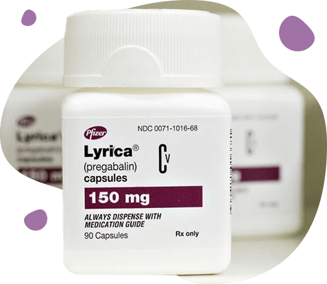
The appearance of the drug gabapentin in the 90s opened up new prospects in the treatment of neuropathic pain and many other chronic pain syndromes. Gabapentin proved to be an effective and safe drug in the treatment of a variety of neuropathic pain syndromes. More recently, in the United States and Europe, a new drug was registered, pregabalin - a continuation of developments in the direction of specific drugs that act independently of the etiology of the central mechanisms of neuropathic pain. It should be emphasized that despite the certain effectiveness of the above-mentioned different groups of drugs, neuropathic pain is not included in the number of indications for use in most of them. The exceptions are: gabapentin and pregabalin, they are registered for the treatment of peripheral and central neuropathic pain, carbamazepine - for the treatment of only trigeminal neuralgia.
Pregabalin is close in its mechanism of action to the well-known drug gabapentin in Russia. However, it has a number of certain differences and significant advantages. Pregabalin is a derivative of gamma-aminobutyric acid and is essentially its analogue. Pregabalin and gabapentin have a similar pharmacological profile. These drugs belong to one class of agents having high affinity for a25-protein of calcium channels in the central nervous system. It is believed that upon modulation of the function of calcium channels (decreasing the entry of calcium into the cell), the release of a number of neurotransmitters (including glutamate, norepinephrine and substance P) in overexcited neurons decreases. As the release of neurotransmitters decreases, the probability of transmission of a nerve impulse to the next neuron becomes lower, which contributes to the reduction of pain. It is important to note that pregabalin causes an effect only under conditions of overexcitation, which manifests itself in modulation, leading to a transition to a normal state. By reducing the release of neurotransmitters, pregabalin selectively inhibits the excitability of a network of neurons, and only in pathological conditions.
Both drugs have been shown to be effective in treating various neuropathic pain. Gabapentin can be taken without regard to meals. No determination of serum concentrations is necessary to optimize treatment. In case of neuropathic pain, the drug should be titrated starting from 300 mg / day, increasing by 300 mg per day, to the target daily dose of 1800 mg. When titrating the dose of the drug, the daily dose cannot be divided into equal parts. If necessary, it can be increased to 3600 mg. The time between doses should not exceed 12 hours. Elderly patients may need a dose adjustment, as they often have impaired renal function. The most common side effects (15%): dizziness (21.1%), drowsiness (16.1%), less frequent diarrhea (5.6%), headache (5.5%), nausea (5.5%) ), peripheral edema (5.4%) and asthenia (5%).
A large number of studies of the effectiveness of pregabalin have been conducted on models of postherpetic neuralgia and diabetic pain neuropathy. Doses of pregabalin from 300 to 600 mg / day have been shown to be most effective compared to placebo, significantly reducing pain and sleep disturbances. The range of daily doses of pregabalin is 150-600 mg / day in 2 or 3 doses. The drug can be taken before, during or after a meal. Available in capsules of 75 mg, 100 mg, 150 mg, 200 mg and 300 mg, capsules of 25 mg and 50 mg for patients with impaired renal function. In the treatment of peripheral neuropathic pain, the starting dose may be 150 mg / day. Depending on the effect and tolerability, the dose can be increased to 300 mg / day after 3-7 days. If necessary, you can increase the dose to a maximum (600 mg / day) after a 7-day interval. In accordance with the experience of using the drug, if necessary, stop taking it is recommended to reduce the dose gradually over the course of a week. Pregabalin is not metabolized in the liver and does not bind to plasma proteins, so it practically does not interact with other drugs. Pregabalin is well tolerated. The most common adverse reactions are dizziness and drowsiness. These adverse events are similar to those most common with gabapentin.
For patients with painful diabetic polyneuropathy, the maximum recommended dose of pregabalin is 100 mg 3 times a day (300 mg / day). Patients with creatinine clearance of 160 ml / min and the elderly should start taking the drug with a dose of 50 mg 3 times a day (150 mg / day) and, depending on the effect and tolerability, the dose during the first week of treatment can be increased to 300 mg / day
For patients with postherpetic neuralgia, the dose of pregabalin is from 75 to 150 mg 2 times a day or from 50 to 100 mg 3 times a day (150-300 mg / day). Patients with creatinine clearance of 160 ml / min and the elderly should start taking the drug with a dose of 75 mg 2 times a day or 50 mg 3 times a day (150 mg / day) and, depending on the effect and tolerance, the dose during the first week treatment may be increased to 300 mg / day.
As a result, taking into account evidence-based studies of the above drugs, the pharmacotherapy algorithm for neuropathic pain can be represented as follows (scheme). Scheme: Algorithm for treating neuropathic pain as part of primary care.
TCAs - tricyclic antidepressants (amntriptyline); SSRIs - selective serotonin reuptake inhibitors (fluoxetine, sertraline, paroxetine, citalopram); SSRIs - selective serotonin and noradrenaline reuptake inhibitors (duloxetine, venlafaxine).
Local treatment with lidocaine, indicated for postherpetic neuralgia and focal neuropathy, can be tested first (transdermal transport system with 5% lidocaine). For neuropathic pain of a different origin, as well as for unsuccessful treatment with lidocaine, we recommend oral monotherapy with gabapentin or pregabalin, a tricyclic antidepressant or a mixed serotonin-noradrenaline reuptake inhibitor. Of these treatments, gabapentin or pregabalin appear to be best tolerated; they are characterized by a very small drug interaction. Tricyclic antidepressants appear to be also effective and less expensive; however, their use is more likely to cause adverse events, and they are relatively contraindicated in serious cardiovascular diseases (electrocardiographic studies are recommended before tricyclic antidepressants are prescribed), orthostatic hypotension, urinary retention, and angle-closure glaucoma. Newer mixed serotonin-noradrenaline reuptake inhibitors (e.g., venlafaxine, duloxetine) are probably less effective than tricyclic antidepressants, but they are better tolerated.
Little is known about whether a reaction to one drug is a predictor of a reaction to another drug. However, if the first tested oral preparation is ineffective or poorly tolerated by the patient, then alternative monotherapy should apparently be used.
If all tested first-line oral monotherapy types are ineffective or poorly tolerated, we recommend that you start monotherapy with tramadol or an opiate analgesic.
When prescribing opiate analgesics for a long time, special requirements must be observed to control the use of these drugs. Tramadol is also available in fixed dose combination with paracetamol. The choice of the maximum dose of tramadol depends on the risk of toxic effects of paracetamol on the liver (i.e., the daily dose of paracetamol should be less than 4000 mg).
 DE
DE FR
FR IT
IT ES
ES
